Sneak up on this brain. You can't.
melanogaster
What you are looking at
Every dot is a real neuron. The dim cloud is 23,210 cell-body positions from the FlyWire connectome -- the first complete wiring map of an adult brain, traced neuron by neuron from electron-microscope images of one fruit fly. The brighter dots are a circuit of of those neurons, connected by of their real synapses, being simulated live on this page -- one thousand steps per second, spike by spike. Nothing is scripted or pre-recorded. When a dot flashes, that cell just fired.
The two large amber dots are the Giant Fibers -- the fly's escape command neurons. One spike from either of them means one thing: takeoff, now.
Why you can't sneak up on it
Your cursor plays the part of a predator. Its motion is converted into input for the circuit's real looming detectors -- the neurons a fly uses to see something rushing toward it. From there, two signals race toward the Giant Fiber: fast electrical excitation from the looming detectors (), and a slower wave of inhibition () that arrives about 4 milliseconds late and vetoes false alarms.
The race is the decision. Creep in slowly and the veto keeps winning -- watch the amber trace get shoved down each time it rises. Lunge, and the excitation outruns its own veto: the trace hits threshold and the Giant Fiber fires within a few milliseconds. Real flies work exactly this way, which is why a flyswatter beats a creeping hand.
How to talk to it
It has no language -- but it has senses, and they are listening. Everything you can say to it, and how it can answer:
| move / lunge | looming input to its LC4 and LPLC2 visual neurons -- approach speed matters, position matters, side matters |
| blow on it | wind onto its real sensory partner neurons, the pathway a fly's antennae use to feel a gust |
| tap the glass | a substrate vibration into the same sensory pathway, sharper and shorter |
| touch a region | clicking the brain stimulates the ~60 nearest neurons directly -- like optogenetics, light-switching cells on |
| tumble it | the cloud is real 3-D anatomy -- every soma sits where it sat in her head. Drag with a mouse to rotate it; the box, origin and axes ride along. On touch, dragging remains the predator |
| slow time down | the sim rate slider runs the identical physics down to 1/1000th speed -- a real trickle. Single spikes become visible, and every line one lights on its way out is a measured synapse: warm lines excite, cool lines inhibit. At full speed only a sampled shimmer is drawn |
| listen | with sound on, each population keeps its own note on a pentatonic scale -- looming detectors ring high, command neurons low, and the Giant Fiber lands as a thud that falls to the tonic. Whatever fires together lands as a chord; slowed to a trickle, every note is one spike |
| its replies | spikes. The transcript translates its command neurons for you: walking, grooming, backing up, darting, escape |
What is real, and what is not
Real: which neurons exist, which connects to which, how many synapses each connection has, which are excitatory and which inhibitory, and where every cell body sits -- all measured from an actual fly brain (FlyWire, FAFB v783).
Modeled: the physics of each neuron (a simple leaky integrate-and-fire unit), the conversion of your cursor into looming, and the plain-language translations in the transcript. The other 99.5% of the fly's brain is not simulated -- its hum is approximated with gentle background noise. So this is not a fly's mind. It is one true reflex of one real animal, running -- the decision to flee you is made by measured synapses, not by code that mentions your cursor.
Connectome data: FlyWire (Dorkenwald et al., Schlegel et al., Nature 2024), CC BY-NC 4.0. Simulation ported from the open-source DesktopFly project; same circuit, same constants, verified against the same test invariants.
The process
how one fly became the most completely known animal that has ever lived
One animal, one morning
Everything in this room comes from a single individual: a female Drosophila melanogaster, laboratory genotype [iso] w1118 × [iso] Canton-S G1, seven days old. At the Howard Hughes Medical Institute's Janelia Research Campus in Virginia, the group of neuroscientist Davi Bock fixed her brain, stained it with heavy metals so that membranes and synapses would catch electrons, and set it in resin.
Then a diamond knife shaved that brain -- about the size of a poppy seed -- into 7,062 slices, each 35–40 nanometres thick: roughly two thousand slices to equal one human hair. The sectioning alone took some three weeks, and 7,050 of the slices survived the knife. That is a 99.8 per cent success rate, and the missing dozen still keep anatomists up at night.
A camera built for one job
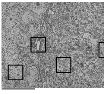
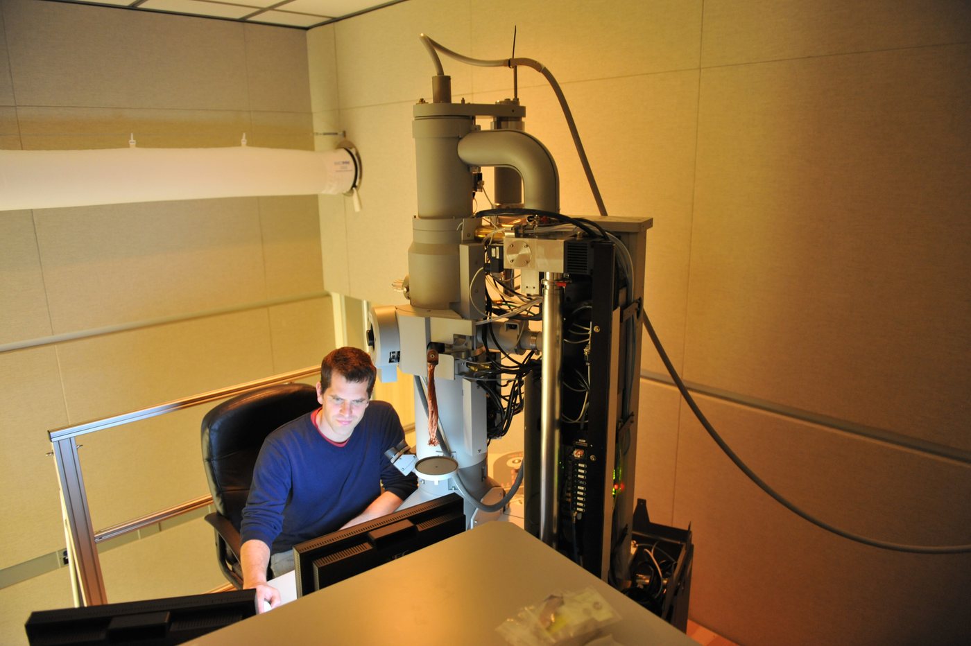
No electron microscope on Earth was fast enough to photograph all of it in a lifetime, so Bock's team built one: TEMCA2, a transmission electron microscope fitted with an array of high-speed cameras and a robotic sample loader, able to image an entire brain slice in under seven minutes at 4 nanometres per pixel.
Running two of these machines produced 21 million images -- about 106 terabytes -- of one poppy seed. Published openly in 2018, the volume was named FAFB: the Full Adult Fly Brain. It was, at the time, the largest complete brain ever imaged at synapse resolution.
The tracing
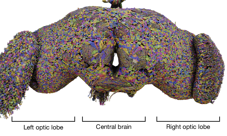
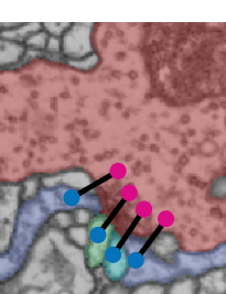
A brain-sized photograph is not a wiring diagram -- someone still has to follow every wire through thousands of slices. At Princeton, the labs of Sebastian Seung and Mala Murthy ran convolutional networks over the volume to segment it into neurons, then opened the whole thing to the world as FlyWire: a browser tool where anyone could proofread the AI's work, wire by wire.
A community from 54 institutions, alongside citizen scientists around the world -- among them veterans of the neuron-tracing game Eyewire -- spent roughly 33 person-years correcting it. Every neuron in this exhibit was checked by human eyes, most of them belonging to volunteers.
The map
On 2 October 2024, the finished connectome was published in Nature: 139,255 neurons, 54.5 million synapses, more than 8,400 cell types -- the first complete wiring diagram of an adult brain of any animal, released free for anyone to study.
Which is why a laurel-forest museum on Tenerife can run a piece of her in your browser. She was never given a name. If it helps, neither is anyone else in the connectome literature -- but nobody else has been read so completely, or remembered so well.
Go deeper: the connectome paper and the whole FlyWire collection in Nature · Codex, the explorer where you can look up any of her 139,255 neurons · FlyWire · the original Janelia story of the imaging. Figures above are cropped from Dorkenwald et al. (2024), used under CC BY 4.0.